Introduction to Animal Cell Diagrams

Animal cell diagram coloring – Animal cell diagrams serve as fundamental visual tools in biology education, providing a simplified yet informative representation of the complex structures and functions within an animal cell. These diagrams are crucial for understanding the intricate organization of cellular components and their interrelationships, which would be significantly more challenging to grasp from textual descriptions alone. Effective visualization is key to unlocking comprehension of cellular processes.Visual aids, such as animal cell diagrams, are essential for bridging the gap between abstract concepts and concrete understanding.
The human brain processes visual information far more efficiently than text, making diagrams particularly effective in conveying the spatial relationships and relative sizes of different organelles within the cell. This facilitates easier memorization and a more intuitive understanding of cell biology.
Common Components of Animal Cell Diagrams
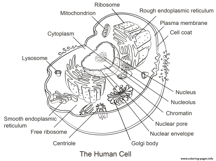
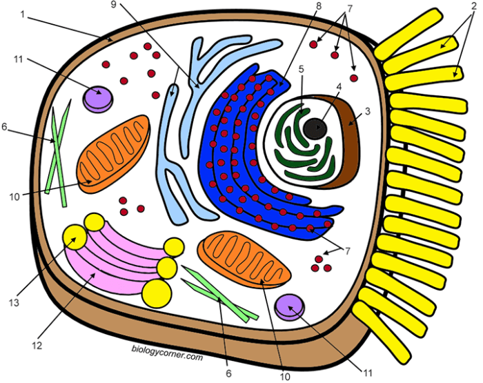
A typical animal cell diagram will illustrate several key organelles and structures. These components, while varying slightly in complexity depending on the diagram’s intended audience and level of detail, usually include a cell membrane, cytoplasm, nucleus, ribosomes, endoplasmic reticulum (both rough and smooth), Golgi apparatus, mitochondria, lysosomes, and sometimes centrioles. The cell membrane, depicted as an outer boundary, represents the selectively permeable barrier regulating the passage of substances into and out of the cell.
The cytoplasm, the jelly-like substance filling the cell, houses the various organelles. The nucleus, often shown as a large, centrally located structure, contains the cell’s genetic material (DNA). Ribosomes, small granular structures, are responsible for protein synthesis. The endoplasmic reticulum (ER), a network of membranes, plays a role in protein and lipid synthesis and transport. The rough ER is studded with ribosomes, while the smooth ER lacks them.
The Golgi apparatus, often depicted as a stack of flattened sacs, modifies and packages proteins for secretion. Mitochondria, often described as the “powerhouses” of the cell, are responsible for generating energy (ATP). Lysosomes contain enzymes for breaking down waste materials. Finally, centrioles, cylindrical structures, play a role in cell division. The relative sizes and positions of these organelles are typically represented to scale, though simplifications are often necessary for clarity.
Key Components of an Animal Cell Diagram

Understanding the intricate workings of an animal cell requires a thorough grasp of its constituent organelles. This section details the major components, their functions, and visual representations commonly used in diagrams. Accurate depiction of these organelles and their relative sizes is crucial for a comprehensive understanding of cellular processes.
Animal cell diagram coloring offers a unique blend of education and creativity, allowing students to visualize complex biological structures. For a different creative outlet, consider checking out some fun designs like those available at anime halloween coloring pages , which offer a vibrant contrast. Returning to the scientific realm, detailed animal cell diagrams provide a valuable learning experience, especially when brought to life with color.
Animal Cell Organelles
The following table summarizes the key organelles found within animal cells, providing functional descriptions and suggestions for visual representation in a diagram. Color choices are subjective and can be adapted based on personal preference or educational context; however, using contrasting colors enhances clarity and memorability.
| Organelle Name | Function | Color Suggestion | Visual Representation |
|---|---|---|---|
| Nucleus | Contains the cell’s genetic material (DNA) and controls cell activities. | Dark Purple | Large, centrally located, often depicted as a darker, round structure with a slightly lighter nucleolus. |
| Ribosomes | Synthesize proteins. | Dark Blue | Small, numerous dots scattered throughout the cytoplasm, possibly clustered on the endoplasmic reticulum. |
| Endoplasmic Reticulum (ER) | Network of membranes involved in protein and lipid synthesis and transport. Rough ER (with ribosomes) and smooth ER (without ribosomes) are distinct. | Light Blue (Rough ER), Light Green (Smooth ER) | Network of interconnected tubes and sacs; rough ER appears rough due to attached ribosomes. |
| Golgi Apparatus (Golgi Body) | Processes, packages, and distributes proteins and lipids. | Yellow | Series of flattened sacs (cisternae) stacked upon each other, often depicted with vesicles budding off. |
| Mitochondria | Generate energy (ATP) through cellular respiration. | Red | Rod-shaped or oval structures, often with internal cristae (folds) represented as inner lines. |
| Lysosomes | Break down waste materials and cellular debris. | Orange | Small, spherical sacs, often depicted as smaller than mitochondria. |
| Cytoplasm | The jelly-like substance filling the cell, containing organelles and cytosol. | Light Pink | Fills the space between the cell membrane and the nucleus. |
| Cell Membrane | Encloses the cell, regulating the passage of substances in and out. | Light Brown | Thin outer boundary of the cell, often represented as a single line. |
| Centrioles (only in animal cells) | Play a role in cell division. | Dark Green | Pair of cylindrical structures, usually located near the nucleus. |
A Text-Based Animal Cell Diagram
Imagine a circle (the cell membrane) approximately 10 units in diameter. Near the center, a large, dark purple circle (the nucleus), about 3 units in diameter, dominates. Within the nucleus, a slightly lighter spot represents the nucleolus. Scattered throughout the cytoplasm (light pink) are numerous small dark blue dots (ribosomes). A network of light blue (rough ER) and light green (smooth ER) tubes and sacs extends throughout the cytoplasm.
Several rod-shaped red structures (mitochondria), about 1.5 units long, are dispersed within the cytoplasm. Smaller orange circles (lysosomes) are scattered amongst the other organelles. A stack of flattened yellow sacs (Golgi apparatus) is located near the nucleus. A pair of dark green cylindrical structures (centrioles) are situated close to the nucleus.
Differences Between Plant and Animal Cells
Plant cells possess several key structures absent in animal cells. These include a rigid cell wall providing structural support and protection, large central vacuoles for storage and turgor pressure regulation, and chloroplasts responsible for photosynthesis. The absence of these structures in animal cells reflects the different lifestyles and metabolic needs of these two cell types. For instance, the rigid cell wall in plants is essential for maintaining their shape and resisting osmotic pressure, while animal cells rely on flexible cell membranes and cytoskeletal support.
The presence of chloroplasts in plants enables them to produce their own food through photosynthesis, a process not undertaken by animal cells, which instead obtain nutrients through consumption.
Educational Applications of Colored Diagrams
Colored diagrams of animal cells offer a powerful pedagogical tool, significantly enhancing understanding and retention of complex biological concepts. The strategic use of color allows for a more intuitive grasp of cellular structures and their interrelationships, transforming a static image into a dynamic learning experience. This is particularly crucial when teaching processes that involve multiple organelles and steps.The visual impact of a color-coded animal cell diagram facilitates comprehension of cellular processes such as respiration and protein synthesis.
By assigning distinct colors to different organelles and molecules involved, the diagram transforms a complex series of reactions into a more easily digestible visual narrative. For example, the mitochondria, the powerhouse of the cell, could be depicted in a vibrant red to highlight its role in energy production, while ribosomes, the protein synthesis sites, might be shown in a contrasting blue to emphasize their distinct function.
This visual differentiation aids in separating and understanding the individual steps of each process.
Illustrating Cellular Processes with Color
A colored diagram effectively demonstrates the intricate dance of molecules during cellular respiration. The movement of glucose through glycolysis, the Krebs cycle within the mitochondrial matrix (represented by a specific shade of red), and the final electron transport chain across the inner mitochondrial membrane (depicted in a different shade of red to show distinct locations) can be visually tracked using color-coding.
Similarly, protein synthesis can be elucidated by highlighting the journey of mRNA from the nucleus (perhaps in a light yellow), its interaction with ribosomes (blue), the role of tRNA (a distinct green), and the eventual translocation of the polypeptide chain (a contrasting purple) to the endoplasmic reticulum (a pale orange). The clear visual distinctions provided by color coding facilitate a deeper understanding of the spatial and temporal aspects of these complex processes.
Enhancing Memory Retention Through Color-Coding
Color-coding significantly improves memory recall. Associating specific colors with particular organelles and their functions creates strong visual mnemonics. For instance, the Golgi apparatus, responsible for packaging and secretion, could be depicted in a bright green, reminding students of its role in “packaging” like green packages. Similarly, lysosomes, responsible for waste breakdown, could be represented in a dark purple, visually associating them with the idea of “dark” or “waste” disposal.
This strategy leverages the brain’s natural predisposition to remember visual information more effectively than purely textual descriptions. The visual cues provided by the color-coded diagram serve as anchor points for recalling the functions of different organelles.
Benefits Across Diverse Learning Environments
Colored animal cell diagrams are versatile tools applicable across various educational settings. In traditional classroom settings, large-format colored diagrams can serve as focal points for group discussions and interactive learning activities. Teachers can use them to guide students through the processes mentioned above, fostering active participation and collaborative learning. In homeschooling environments, these diagrams offer a flexible and engaging learning resource, allowing for self-paced learning and individualized instruction.
The visual nature of the diagrams makes them particularly beneficial for students with diverse learning styles, including visual learners who benefit greatly from pictorial representations of abstract concepts. The ease of reproduction and accessibility of these diagrams makes them a valuable tool for both in-person and remote learning scenarios.
Creating a Detailed Animal Cell Diagram: Animal Cell Diagram Coloring
Creating a highly detailed and accurate animal cell diagram for a coloring page requires a thorough understanding of the cell’s intricate structure and the function of its various organelles. The challenge lies not only in depicting the organelles correctly but also in maintaining realistic size ratios within the confines of a visually appealing and educational coloring page. This section will delve into the specifics of each organelle and the considerations necessary for creating a truly informative and aesthetically pleasing diagram.
Organelle Descriptions for a Detailed Diagram, Animal cell diagram coloring
The accuracy and completeness of the organelle representations are crucial for a successful educational coloring page. Each organelle should be depicted with its characteristic shape and location within the cell, providing a visual representation of its function.
The Cell Membrane is a selectively permeable barrier surrounding the cell, regulating the passage of substances in and out. It’s a fluid mosaic of lipids and proteins, often depicted as a thin, continuous line in diagrams, but its complex structure should be hinted at through textural details or shading in a coloring page.
The Cytoplasm fills the cell’s interior, a gel-like substance containing various organelles. It should be depicted as a background space, with organelles appropriately spaced within. Consider using a gradient of color to represent the varying density of the cytoplasm.
The Nucleus, the cell’s control center, houses the genetic material (DNA). It’s typically large and spherical, containing a nucleolus (a smaller, darker region involved in ribosome synthesis). The nuclear membrane, with its pores, should be clearly shown as a double membrane surrounding the nucleus.
Ribosomes, the protein synthesis factories, can be free-floating in the cytoplasm or attached to the endoplasmic reticulum. They are tiny and numerous, best represented as small dots or stippled areas, strategically placed throughout the cytoplasm and along the ER.
The Endoplasmic Reticulum (ER) is a network of membranes extending throughout the cytoplasm. The rough ER (studded with ribosomes) should be depicted differently from the smooth ER (lacking ribosomes). The rough ER could be shown as a network of membranes with attached dots (ribosomes), while the smooth ER could be shown as a network of smooth membranes.
The Golgi Apparatus (Golgi Body) modifies, sorts, and packages proteins and lipids. It’s often depicted as a stack of flattened sacs (cisternae). These sacs should be shown as parallel, slightly curved structures.
Mitochondria, the “powerhouses” of the cell, generate energy through cellular respiration. They are often sausage-shaped or oval, with a folded inner membrane (cristae). Their double-membrane structure should be clearly visible.
Lysosomes, containing digestive enzymes, break down waste materials and cellular debris. They are typically depicted as small, membrane-bound vesicles. Their size relative to other organelles should be considered.
Centrioles, involved in cell division, are usually paired cylindrical structures located near the nucleus. They should be shown as small, paired cylinders positioned appropriately near the nucleus.
Challenges in Representing Organelle Size and Shape
Accurately representing the size and shape of organelles in a diagram presents several challenges. Organelles are microscopic, and their actual sizes vary greatly. For instance, the nucleus is significantly larger than a ribosome. Maintaining accurate proportions while also creating a visually appealing and easily understandable diagram requires careful planning and scaling. The three-dimensional nature of organelles also poses a challenge when translating them into a two-dimensional representation.
Importance of Maintaining Accurate Proportions
Maintaining accurate proportions in a detailed animal cell diagram is crucial for its educational value. An inaccurate representation can lead to misconceptions about the relative sizes and spatial relationships of organelles. For example, if the mitochondria are depicted as being the same size as the nucleus, it misrepresents their actual size difference and their respective roles within the cell.
Consistent scaling throughout the diagram ensures that learners develop a correct understanding of the cell’s internal organization. A well-proportioned diagram facilitates a more accurate and effective learning experience.
Commonly Asked Questions
What are the best coloring tools for an animal cell diagram?
Colored pencils, markers, or crayons all work well. The choice depends on personal preference and desired level of detail.
How can I ensure my coloring is accurate and reflects the organelles’ functions?
Use a color key or legend. Consistent color choices for similar functions will enhance understanding.
Are there online resources that can help with animal cell diagram coloring?
Yes, many websites offer printable diagrams and information on cell structures.
Why is accurate representation of size and shape important in an animal cell diagram?
Accurate proportions help to visualize the spatial relationships between organelles and understand their relative importance.


