Cellular Structures

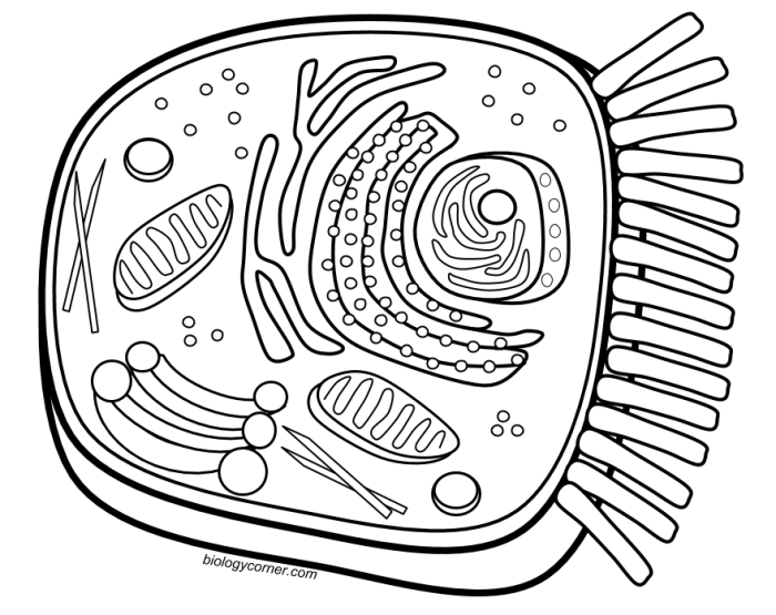
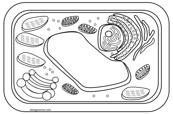
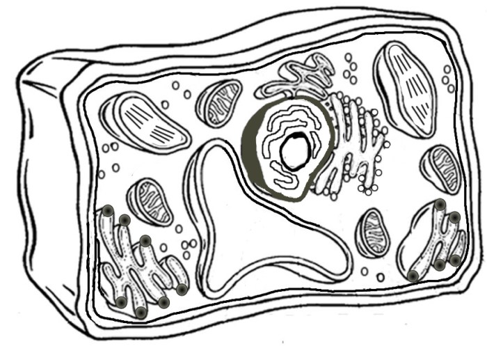
Coloring the cell animal and plant – Okay, so you wanna know about plant and animal cells? Think of it like this: they’re both like tiny cities, but one’s got a sweet suburban vibe, and the other’s more of a bustling metropolis. Let’s break down the differences, Hollywood style!
Plant and Animal Cell Comparison, Coloring the cell animal and plant
Here’s the lowdown on the key differences. We’re talking about the big players—cell wall, chloroplasts, vacuoles, and overall shape. It’s like comparing a perfectly manicured lawn (plant cell) to a wild, energetic party (animal cell).
| Feature | Plant Cell | Animal Cell |
|---|---|---|
| Cell Wall | Present; rigid, made of cellulose; provides support and protection. Think of it as the sturdy brick walls of a castle. | Absent; flexible cell membrane provides support. More like a flexible, breezy beach house. |
| Chloroplasts | Present; conduct photosynthesis, converting sunlight into energy (the plant’s solar panels!). They look like little green ovals, like tiny, energy-producing power plants. | Absent; obtain energy from consuming other organisms. They rely on outside sources for energy, like ordering takeout. |
| Vacuoles | Large central vacuole; stores water, nutrients, and waste. It’s the plant cell’s main storage facility, a huge water tank. | Smaller vacuoles; various functions, including storage. They’re more like smaller, more numerous storage containers, each with specific jobs. |
| Cell Shape | Typically rectangular or cubic due to the rigid cell wall. Think perfectly stacked Lego bricks. | Variable; often round or irregular. More like amoebas, constantly changing shape. |
Typical Plant Cell Structure and Function
Imagine a plant cell as a well-organized factory. The powerhouse is the mitochondria, oval-shaped organelles that generate energy through cellular respiration – they’re like the generators of the factory, providing power for all operations. The chloroplasts, those green ovals, are the solar panels, converting sunlight into energy via photosynthesis. The nucleus, a large, round structure, is the control center, containing the cell’s DNA – the blueprints for everything that happens in the cell.
The endoplasmic reticulum (ER), a network of membranes, is like the factory’s conveyor belt system, transporting materials. The Golgi apparatus, a stack of flattened sacs, is the packaging and shipping department, modifying and exporting proteins. The cell wall, that rigid outer layer, is the factory’s sturdy exterior walls. Finally, the large central vacuole acts as the factory’s warehouse, storing water, nutrients, and waste.
Typical Animal Cell Structure and Function
Now, picture an animal cell as a bustling city. The nucleus, again a round structure, is the city hall, controlling all cellular activities. The mitochondria, those energy powerhouses, are like the city’s power plants, generating energy. The endoplasmic reticulum (ER) is the city’s extensive road network, transporting materials. The Golgi apparatus acts as the city’s postal service, packaging and delivering proteins.
The lysosomes, small, round organelles, are the city’s waste management system, breaking down waste materials. The ribosomes, tiny dots scattered throughout the cell, are the city’s construction workers, synthesizing proteins. The cell membrane, a flexible outer boundary, is the city’s protective wall, regulating what enters and exits.
Relationship Between Cellular Structure and Function
The differences in cellular structure directly reflect the different functions of plant and animal cells. Plant cells, with their cell walls and chloroplasts, are designed for photosynthesis and structural support, allowing them to create their own food and stand tall. Animal cells, lacking these structures, rely on consuming other organisms for energy and have more flexible structures enabling movement and diverse functions.
It’s like comparing a marathon runner (plant cell, steady and strong) to a gymnast (animal cell, agile and adaptable). Each is built for a different purpose, with structures perfectly suited to their respective lifestyles.
Cell Coloring Techniques and Materials: Coloring The Cell Animal And Plant

Alright, peeps! We’ve already nailed down the basics of cell structure – think of it as the foundation of a killer house. Now, let’s talk about thedecor*. We’re going to pimp those cells with some serious color, making them Instagram-worthy under the microscope. Getting a clear view of these tiny titans requires some serious staining techniques, and we’re about to break it all down.
Microscopic Staining Methods for Plant and Animal Cells
Choosing the right staining method is like picking the perfect filter for your selfie – it totally changes the vibe! Different stains highlight different cellular components, giving you a deeper understanding of what’s going on inside those microscopic worlds. Here are some of the hottest staining techniques used by cell biologists:
- Simple Staining: This is your basic, go-to method. It uses a single stain to highlight the overall shape and structure of the cells. Think of it as the “no-makeup makeup” look for your cells – natural but enhanced.
- Differential Staining: This is where things get interesting. Differential staining uses multiple stains to distinguish between different types of cells or cellular components. It’s like adding a pop of color to your outfit – it makes a statement!
- Gram Staining (for bacteria, but relevant for comparison): While not directly used for plant and animal cells, Gram staining is a crucial differential staining technique in microbiology. It distinguishes between Gram-positive and Gram-negative bacteria based on their cell wall composition, resulting in purple or pink coloration respectively. This exemplifies the power of differential staining to reveal crucial differences.
- Negative Staining: Instead of staining the cells directly, this method stains the background, making the cells stand out like rockstars against a dark backdrop. It’s all about contrast!
Comparison of Common Cell Stains
Let’s dive into the specifics of some popular stains and see what they bring to the party. Think of this as your stain cheat sheet for microscopic success!
| Stain Type | Target Components | Resulting Color | Notes |
|---|---|---|---|
| Methylene Blue | Nucleic acids, cytoplasm | Blue | A classic simple stain; relatively inexpensive and easy to use. |
| Iodine | Cellulose (plant cells), starch | Brownish-yellow to dark blue/black (depending on starch presence) | Highlights cell walls in plant cells and reveals the presence of starch granules. |
| Crystal Violet | Cell walls (especially effective in bacteria, but also useful for plant cells) | Purple | Often used in conjunction with other stains as part of differential staining techniques. |
Sample Preparation Before Staining: The Pre-Game Ritual
Before you unleash the staining power, proper sample preparation is crucial. It’s like prepping your canvas before painting a masterpiece. You wouldn’t start painting on a dirty canvas, right? Here’s the lowdown:For both plant and animal cells, the process generally involves creating a thin, even smear or mount on a microscope slide. For plant cells, this might involve gently scraping a bit of tissue from a leaf or stem and then carefully spreading it on the slide.
Okay, so like, coloring those cell diagrams of animals and plants is, kinda, low-key boring, right? But if you need a break, check out these totally adorable cute animal coloring pages printable – they’re, like, way more fun! Then, you can totally get back to those cells with fresh eyes and a happier vibe. It’s a total brain break, you know?
Animal cells, often obtained from cheek swabs or blood samples, require similar careful spreading to ensure a single-cell layer for optimal visualization. This prevents overlapping cells that would obscure details. The use of a coverslip also ensures a flat, even surface for observation. Proper preparation guarantees you won’t miss those tiny details!
Advanced Cell Staining Techniques

Okay, so we’ve covered the basics of cell coloring – think of it like using Crayola crayons on your microscopic masterpieces. But to really level up your cell-visualization game, you need to ditch the crayons and embrace some seriously high-tech staining techniques. These methods allow us to see cellular structures and processes with way more detail and precision than basic dyes ever could.
We’re talking about seeing the inner workings of a cell like never before – think high-definition, microscopic reality TV!Advanced staining techniques offer a major upgrade from basic dyes, providing significantly enhanced resolution and specificity in visualizing cellular components. This allows researchers to gain deeper insights into cellular structure and function. They’re like the difference between watching a grainy VHS tape and streaming in glorious 4K.
Advanced Staining Techniques: A Lineup of Cellular Superstars
Let’s break down some of the awesome advanced staining techniques that are revolutionizing cell biology. These aren’t your grandma’s basic dyes; these are the power players of the cellular world.
- Immunofluorescence (IF): Imagine this: you’ve got a specific cellular protein you want to see, like a hidden celebrity at a party. Immunofluorescence is like having a super-powered spotlight that only shines on that one protein. It uses antibodies (think tiny, protein-seeking missiles) tagged with fluorescent dyes to locate and highlight the target protein within the cell. It’s like giving your protein a neon glow-up! The resulting images show the precise location and distribution of the protein, providing incredible detail about its function and interactions.
- Fluorescent Protein Tagging: This technique is like giving your protein a built-in, glowing GPS tracker. Researchers genetically engineer cells to produce proteins fused with fluorescent proteins like Green Fluorescent Protein (GFP). These proteins naturally glow under specific wavelengths of light, allowing researchers to track the protein’s movement, localization, and interactions within the living cell in real-time. Think of it as a live-action, microscopic movie of your protein’s adventures.
Basic Dyes vs. Advanced Staining Techniques: A Head-to-Head Showdown
Basic dyes are like the trusty sidekick – simple, readily available, and good for a general overview. But advanced techniques? They’re the main event.
| Feature | Basic Dyes | Advanced Staining Techniques (e.g., Immunofluorescence, Fluorescent Protein Tagging) |
|---|---|---|
| Specificity | Low; stains many cellular components non-specifically | High; targets specific molecules or structures |
| Resolution | Limited; provides general morphology | High; allows for detailed visualization of specific structures and processes |
| Cost | Low | High; requires specialized equipment and reagents |
| Complexity | Simple; easy to perform | Complex; requires specialized training and expertise |
| Live Cell Imaging | Generally not suitable | Often possible (especially with fluorescent protein tagging) |
Enhanced Visualization and Understanding of Cellular Processes
These advanced techniques are game-changers for understanding cellular processes. For example, immunofluorescence allows researchers to visualize the location of specific proteins involved in cell signaling pathways, providing crucial insights into how cells communicate and respond to their environment. Fluorescent protein tagging allows for real-time tracking of protein movement and interactions, providing a dynamic view of cellular processes like protein trafficking or chromosome segregation during cell division.
Think of it as having a backstage pass to the cell’s most exciting events! This level of detail is crucial for understanding everything from disease mechanisms to the development of new therapies. For instance, researchers studying cancer can use these techniques to visualize the distribution of cancer-related proteins within tumor cells, providing valuable information for developing targeted therapies.
It’s like having X-ray vision for the cellular world.
Common Queries
What are some safety precautions when working with cell stains?
Always wear appropriate personal protective equipment (PPE), including gloves and eye protection. Work in a well-ventilated area and follow the manufacturer’s instructions for handling and disposal of stains.
Can I use household dyes to stain cells?
No, household dyes are not suitable for staining cells for microscopic observation. They lack the specific properties needed to bind to cellular components and provide clear visualization.
How do I choose the right stain for my experiment?
The choice of stain depends on the specific cellular components you want to visualize. Methylene blue stains nuclei, while iodine stains starch granules. Consult scientific literature or staining protocols for guidance.
What is the resolution limit of a light microscope when observing stained cells?
The resolution limit of a light microscope is approximately 200 nm. This means that structures smaller than this cannot be clearly resolved, even with staining.


